
Hemodynamic Modeling of Branch Retinal Artery Occlusion Involving Asymmetric Vascular Stenosis
Article Sidebar
Main Article Content
 https://orcid.org/0000-0003-4297-1721
Abstract
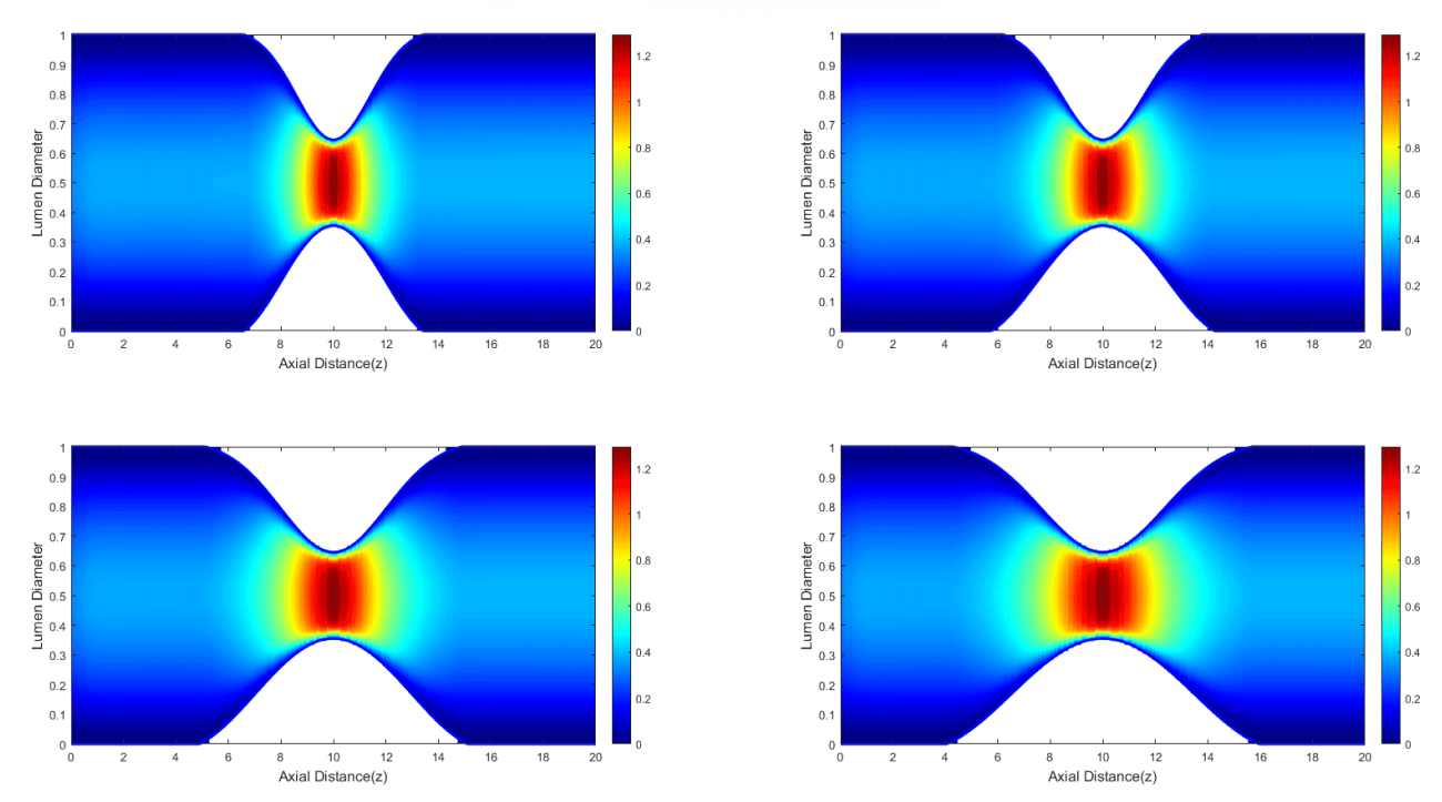
The narrowing of retinal blood vessels, especially in conditions involving branch retinal artery occlusion (BRAO), can cause gradual and painless vision deterioration. Such vascular obstruction is also a contributing factor to eye stroke. This study investigates the influence of three asymmetric stenosis geometries, namely Bell--Cosine, Cosine--Overlapping, and Bell--Overlapping, on fluid flow characterized as Newtonian, incompressible, and steady. The mathematical formulation is derived from the Navier--Stokes equations, discretized using the Finite Volume Method (FVM) and solved through the Semi-Implicit Method for Pressure-Linked Equations (SIMPLE) algorithm. Numerical simulations are performed in MATLAB with variations in stenosis length. The results demonstrate how different geometric shapes and stenosis lengths affect blood flow rate and pressure distribution. Among all configurations, the Bell--Cosine geometry consistently produces a flow rate above the normal threshold and a pressure level below the normal range for each stenosis length ($40\,\mu\text{m}$, $50\,\mu\text{m}$, $60\,\mu\text{m}$, $70\,\mu\text{m}$), compared with the other geometries. For every geometric arrangement, stenosis length plays a role in altering the flow behavior and pressure field around the constriction, while the peak velocity and peak pressure remain essentially unchanged.
Article Details

This work is licensed under a Creative Commons Attribution-NonCommercial 4.0 International License.
References
[1] Biehler R, Scholz RW, Straßer R, Winkelmann B. Didactics of Mathematics as a Scientific Discipline. Dordrecht: Kluwer Academic Publishers; 2002. doi:10.1007/0-306-47204-x.
[2] Versteeg MW H K. An introduction to computational fluid dynamics: The finite volume method (2nd ed.). vol. M. 2nd ed. Harlow, England: Pearson Education; 2007.
[3] Lu Y, Bernabeu MO, Lammer J, Cai CC, Jones ML, Franco CA, et al. Computational fluid dynamics assisted characteri- zation of parafoveal hemodynamics in normal and diabetic eyes using adaptive optics scanning laser ophthalmoscopy. Biomedical Optics Express. 2016 dec;7(12):4958. doi:10.1364/boe.7.004958.
[4] Ong CW, Tan B, Hussain S, Chuangsuwanich T, Braeu FA, Cui F. Evaluation of the effect of hemodynamic factors on retinal microcirculation by using 3D confocal image-based computational fluid dynamics. Frontiers in Bioengineering and Biotechnology. 2024 nov;12. doi:10.3389/fbioe.2024.1489172.
[5] Bénard-Séguin É, Nahab F, Pendley AM, Rodriguez Duran M, Torres Soto M, Keadey M, et al. Eye stroke pro- tocol in in the emergency department. Journal of Stroke and Cerebrovascular Diseases. 2024;33(9):107895. doi:10.1016/j.jstrokecerebrovasdis.2024.107895.
[6] Genevois O, Paques M, Simonutti M, Sercombe R, Seylaz J, Gaudric A, et al. Microvascular Remodeling after Occlusion- Recanalization of a Branch Retinal Vein in Rats. Investigative Ophthalmology and Visual Science. 2004 feb;45(2):594-600. doi:10.1167/iovs.03-0764.
[7] Mason JO, Shah AA, Vail RS, Nixon PA, Ready EL, Kimble JA. Branch Retinal Artery Occlusion: Visual Prognosis. American Journal of Ophthalmology. 2008;146(3):455-7. doi:10.1016/j.ajo.2008.05.009.
[8] Toolan KJ, Minaker S, Civantos J. Branch retinal artery occlusion (BRAO) after macroaneurysm. American Journal of Ophthalmology Case Reports. 2024;36:102108. doi:10.1016/j.ajoc.2024.102108.
[9] Fatahillah A, Widodo B, Roslan R. The Modeling and Numerical Solution of Branch Retinal Artery Occlusion. Malaysian Journal of Mathematical Sciences. 2025;19(1):1-16. doi:10.47836/mjms.19.1.01.
[10] Kumar S, Kumar S. Blood Flow with Heat Transfer through Different Geometries of Stenotic Arteries. Trends in Sciences. 2023;20(11):6965. doi:10.48048/tis.2023.6965.
[11] Fatahillah A, Widodo B, Roslan R. Mathematical Modeling and Numerical Simulation of Central Retinal Artery Occlu-sion (CRAO) with Asymmetric Stenosis. Statistics, Optimization and Information Computing. 2025;13(5):2079-104. doi:10.19139/soic-2310-5070-2381.
[12] Aruchamy P, Mahagaonkar P, Ganesan G, Dhandapani PB, Yahya NI. Identifying the Fetal Heart Rate and Gender with Intuitionistic Fuzzy Total Edge Magic Labelling. Jambura Journal of Biomathematics (JJBM). 2025;6(2):159-65. doi:10.37905/jjbm.v6i2.30951.
[13] Das K, Kumar GR, Ramesh K, Biswas MHA. A Qualitative Analysis of Leukemia Fractional Order SICW Model. Jambura Journal of Biomathematics (JJBM). 2024;5(1):46-53. doi:10.37905/jjbm.v5i1.24961.
[14] Chung TJ. Computational Fluid Dynamics. Cambridge University Press; 2002. doi:10.1017/CBO9780511606205. [15] Guidoboni G, Harris A, Carichino L, Arieli Y, Siesky BA. Effect of intraocular pressure on the hemodynamics of the central retinal artery: A mathematical model. Mathematical Biosciences and Engineering. 2014;11(3):523-46. doi:10.3934/mbe.2014.11.523.
[16] Wu LT, Wang JL, Wang YL. Ophthalmic artery morphological and hemodynamic features in acute coronary syndrome. Investigative Ophthalmology and Visual Science. 2021;62(14):7. doi:10.1167/iovs.62.14.7.
[17] Hua Zhou Z, Ru Cheng X, Xin Guan J, Zhao L, ling Wang Y, lin Wang J. A Nomogram Based on Oc- ular Hemodynamics for Predicting Ischemic Stroke. American Journal of Ophthalmology. 2025;274:91-100. doi:10.1016/j.ajo.2025.02.034.
[18] Kim HM, Woo SJ. Clinical characteristics of recurrent non-arteritic retinal artery occlusion. BMJ Open Ophthalmology. 2024;9(1):e001636. doi:10.1136/bmjophth-2024-001636.
[19] Ismail A, Chen HC, Faye I, Tang TB. Longitudinal effects of common carotid artery stenosis on ocular hemodynamics assessed using laser speckle flowgraphy in a rabbit model. Scientific Reports. 2020;10(1):15829. doi:10.1038/s41598-020-72556-9.
[20] Liu WL, Wu LT, Wang JL, Sun J, Cheng XR, Zhou ZH, et al. Effect of PCI on ophthalmic artery hemodynamics in patients with acute coronary syndrome. Frontiers in Medicine. 2024;11:1367900. doi:10.3389/fmed.2024.1367900.
[21] De Salvo G, Oshallah M, Sepetis AE, Borbara R, Oliverio GW, Meduri A, et al. Inner Retinal Thinning Compari- son between Branch Retinal Artery Occlusion and Primary Open-Angle Glaucoma. Diagnostics. 2023;13(22):3428. doi:10.3390/diagnostics13223428.
[22] Goldenberg D, Shahar J, Loewenstein A, Goldstein M. Diameters of retinal blood vessels in a healthy cohort as measured by spectral domain optical coherence tomography. Retina. 2013;33(9):1888-94. doi:10.1097/IAE.0b013e31829477f2.
[23] Ding Y, Jiang Z, Jiang J, Yang G, Li Y, Tao L. Computational assessment of blood lipid influence on hemodynamics in human retinal vessels. Scientific Reports. 2025;15(1):16542. doi:10.1038/s41598-025-98767-6.
[24] Hayreh SS. Acute retinal arterial occlusive disorders. Progress in Retinal and Eye Research. 2011;30(5):359-94. doi:10.1016/j.preteyeres.2011.05.001.
[25] Yu DY, Cringle SJ, Balaratnasingam C, Morgan WH, Yu PK, Su EN. Retinal ganglion cells: Energetics, compart- mentation, axonal transport, cytoskeletons and vulnerability. Progress in Retinal and Eye Research. 2013;36:217-46. doi:10.1016/j.preteyeres.2013.07.001.